 Tweet
Tweet
I'm kinda a handy-man and have several hobbies, mostly focused on metalworking.
At some point I bougth a Amscope stereo microscope for working on intrincate small mechanism and inspection, and despite the cheap price, looking intro the eyepieces at anythig was WOW!, and I have been looking at everithing on hand from then on (insecs, dirt, plants, etc...), also I got very interested in optical measure of length.
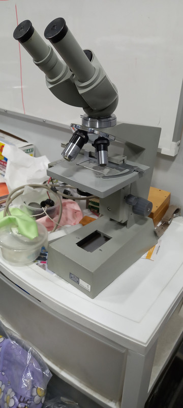
Now, I got to a antique store and see a dusty and forgotten light microscope, I recognice the brand Carl Zeiss Jena (as I own a couple of metrology instruments from that brand, and they are good) and got it dirt cheap. The microscope is complete, but full of dust and rust, all optics are fogged and dusty, and the electrical parts have been intervened and destroyed.
After some research, the microscope is a Laboval 2, seems a routine microscope for the 70?s. I have not been able to find the owner manual or service manual, or any information at all exept for some mechanical dissasembly of the fine focus system.
It came with all original Carl Zeiss Jena parts:
- two A10X 14 Eyepieces
Objectives:
- 3.2 / 0.10 Semiplan
- 10 / 0.25
- 40 / 0,65
- HI 100 / 1,25 oil inmersion with a iris??
Also it has a 1,6X binoviewer. It seems that with the 40x objective the magnification (40 x 1,6 x 10 = 640 ?) its almost getting intro empy magnification treshold?? the same with the 100X. not sure
For a couple of weeks I have been working on it, cleaning and bringing it to fully mechanical function (never have seen grease goin so hard!) and also optical cleaniness. Now I can use it but the lamp housing and electrics still need some work.
Using the cellphone flashlight on top of the base for ilumination, I put a drop of a rain water pond on top of a slide and I was able to see very small dots revoling on it, very impresive! pushing the magnification I see these are microorganism with I could not focus properly because of the very shallow deep of field, or maybe still fogged and dusty optics (inside the binoviewer witch I haven't been able to disassembly), or the very poor ligth source and optical misligment of components after all the disassembly, I dont now, but I hope to solve it!
Many thans in advance for your advise and teachings.
Best Regards.
At some point I bougth a Amscope stereo microscope for working on intrincate small mechanism and inspection, and despite the cheap price, looking intro the eyepieces at anythig was WOW!, and I have been looking at everithing on hand from then on (insecs, dirt, plants, etc...), also I got very interested in optical measure of length.
Now, I got to a antique store and see a dusty and forgotten light microscope, I recognice the brand Carl Zeiss Jena (as I own a couple of metrology instruments from that brand, and they are good) and got it dirt cheap. The microscope is complete, but full of dust and rust, all optics are fogged and dusty, and the electrical parts have been intervened and destroyed.
After some research, the microscope is a Laboval 2, seems a routine microscope for the 70?s. I have not been able to find the owner manual or service manual, or any information at all exept for some mechanical dissasembly of the fine focus system.
It came with all original Carl Zeiss Jena parts:
- two A10X 14 Eyepieces
Objectives:
- 3.2 / 0.10 Semiplan
- 10 / 0.25
- 40 / 0,65
- HI 100 / 1,25 oil inmersion with a iris??
Also it has a 1,6X binoviewer. It seems that with the 40x objective the magnification (40 x 1,6 x 10 = 640 ?) its almost getting intro empy magnification treshold?? the same with the 100X. not sure
For a couple of weeks I have been working on it, cleaning and bringing it to fully mechanical function (never have seen grease goin so hard!) and also optical cleaniness. Now I can use it but the lamp housing and electrics still need some work.
Using the cellphone flashlight on top of the base for ilumination, I put a drop of a rain water pond on top of a slide and I was able to see very small dots revoling on it, very impresive! pushing the magnification I see these are microorganism with I could not focus properly because of the very shallow deep of field, or maybe still fogged and dusty optics (inside the binoviewer witch I haven't been able to disassembly), or the very poor ligth source and optical misligment of components after all the disassembly, I dont now, but I hope to solve it!
Many thans in advance for your advise and teachings.
Best Regards.
Comment